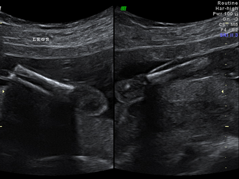
Early Pregnancy
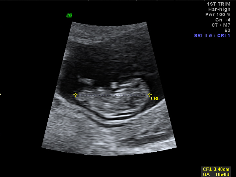
Performed in the first Trimester (4 – 12) weeks of Pregnancy. This examination is usually performed to confirm viability of the pregnancy, confirm dating of the pregnancy and to confirm the number of babies.
In the early stages of the pregnancy, before 7 weeks, a transvaginal ultrasound examination is usually required to see the pregnancy sac in the uterus. Fetal heart activity will usually be found between 5-6 weeks gestation on transvaginal ultrasound.
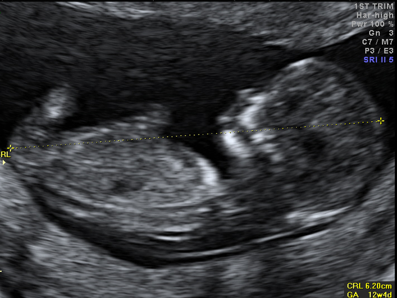
It is important at this stage to confirm that the pregnancy is located in the uterus. Accurate dating of the pregnancy can be undertaken by measurement of the crown rump length of the baby (top to bottom).
This is very accurate within a few days to predict the due date of delivery.
An early examination of a twin or multiple pregnancy can be extremely important to determine whether the twins or multiple babies have come from one or several eggs. This can be very helpful in planning the management of the pregnancy.
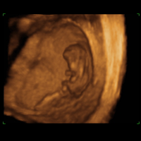
Nuchal Translucency
This is a simple non invasive test best performed between 12-14 weeks of pregnancy to assess the risk of having a baby with a chromosome abnormality.
This test allows up to 95% of babies with Down syndrome and other abnormalities to be identified. This test uses a combination of a scan and a blood test and must be performed in an accredited Fetal medicine centre.
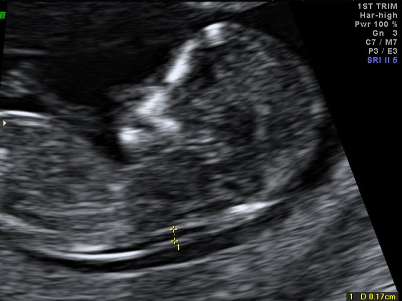
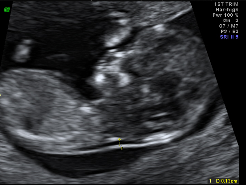
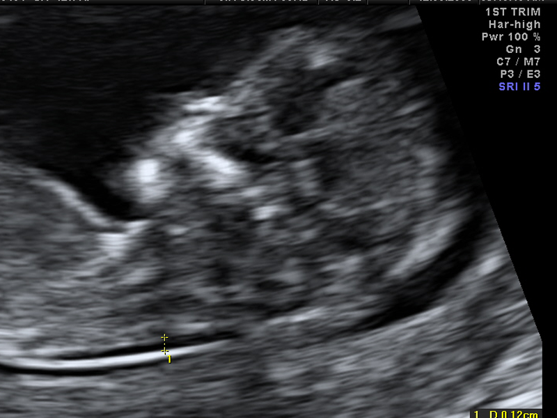
How is the test done? A transabdominal ultrasound examination measuring the thickness of a layer of fluid at the back of the baby’s neck is performed. The presence of the nasal bone is also assessed as are the baby’s heart valves. This helps to make the test more accurate. Some patients may need a transvaginal examination to allow this measurement to be done correctly. This test is not an invasive procedure for your baby and does not carry any risk of miscarriage.
The blood test: The blood test measures PAPP-A and free BHCG, two pregnancy related proteins, produced by the placenta and must be tested between 10 and 13 weeks of pregnancy.
How does the test assess the risk of abnormality? Studies of over 100,000 pregnancies in women of all ages have shown that up to 95% of babies with Down syndrome or other abnormalities can be identified. A special computer program is necessary to calculate this risk and takes into account the results of the blood test, NT measurement, the mother’s age, the stage of pregnancy and any previous baby born with a chromosome abnormality.
What does a high risk result test mean? A high risk result does not automatically mean that a baby is abnormal. A risk greater than 1 in 300 is considered high and this may occur in about 1 in 20 patients. Further testing such as CVS (Chorion Villus Sampling) done at 12-14 weeks or amniocentesis done at 15-16 weeks is usually recommended. The NIPT can also be used to assess the risk of chromosome abnormality in the baby.
A high risk test result, in a baby with normal chromosomes, can indicate an increased risk of a fetal heart abnormality and concerns with the growth of the baby during the pregnancy. Careful assessment of the baby is usually recommended at 16 and 20 weeks of pregnancy.
What does a low risk result test mean? A risk of lower than 1 in 300 suggests a low chance of chromosome abnormality for your baby but this DOES NOT mean no risk.
18-20 Week Morphology
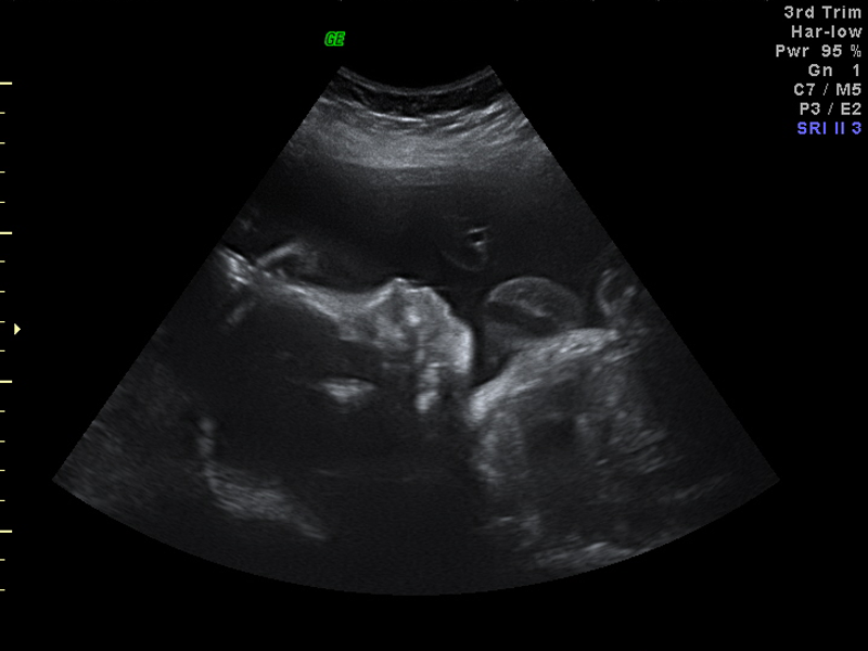
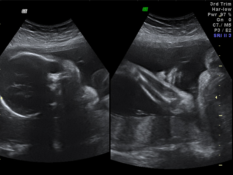
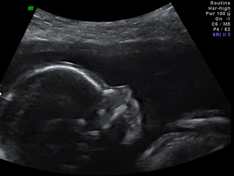
3rd Trimester
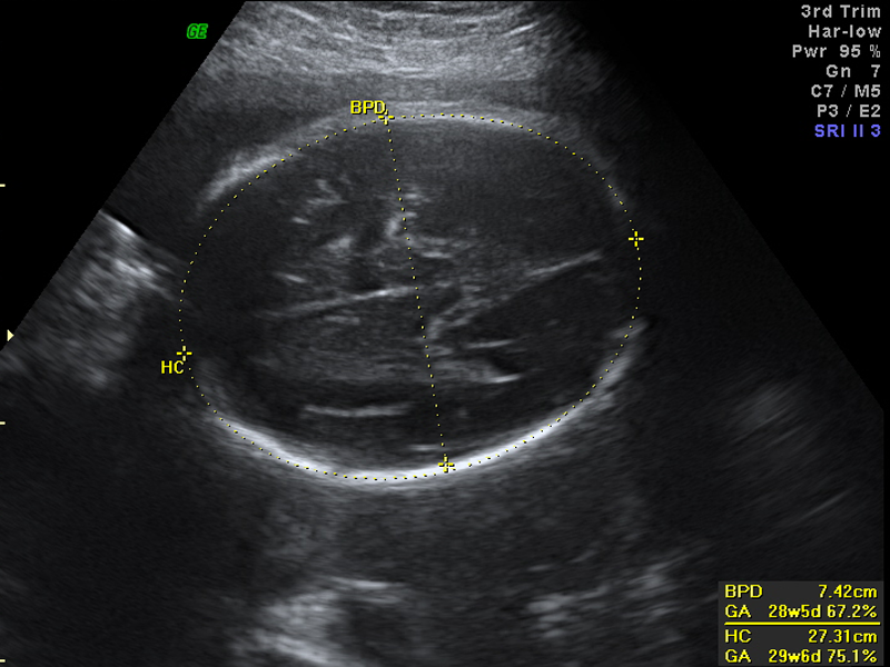
The purpose of this examination is usually to assess the position, growth and wellbeing of the baby. This is often undertaken if there is a concern about the baby’s growth or if the mother has become affected by diabetes or blood pressure. This examination is often repeated several times to assess the growth and wellbeing of each baby in a multiple pregnancy.
During this examination the measurements are obtained of the baby to assess the growth and estimate the fetal weight. The size of the baby can be compared to the size expected for this stage of pregnancy, assessing how well the baby is growing.
Careful examination of the placenta, measurement of the blood flow in the cord with Doppler Ultrasound and assessment of the liquor volume make up the biophysical profile. This can give a very good assessment of how well the baby is or if the baby is at risk of further problems.
The position and presentation (which part of the baby is coming first) of the baby is important at this stage as it can affect the mode of delivery.
The position of the placenta is also assessed because a low position may make a caesarean section necessary for the safe delivery of the baby.
Ectopic Pregnancy
Occasionally a pregnancy may occur outside the uterus; either in the tubes or on the ovary. This may be more common in patients undergoing IVF or who have had recurrent miscarriages
This is known as an ectopic pregnancy and can be life threatening to the mother. This can be detected by an early ultrasound examination performed transvaginally before 6-7 weeks of pregnancy.
Early medical or surgical treatment can then be urgently considered and performed to avoid severe complications to the mother’s health and also preserve her fertility.
3D/4D Ultrasound
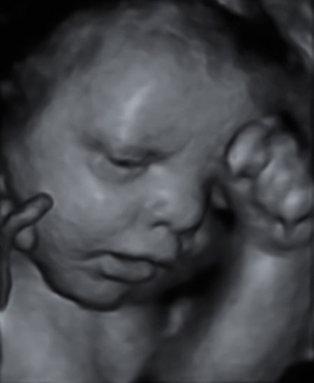
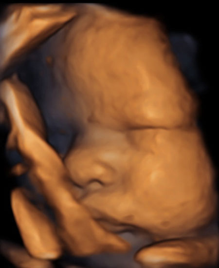
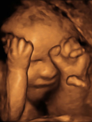
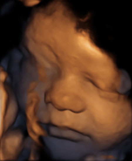
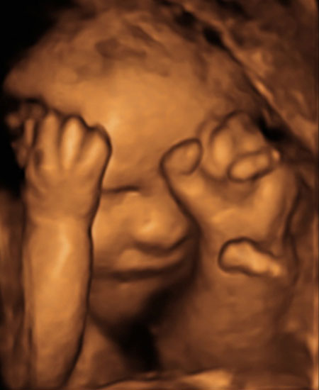
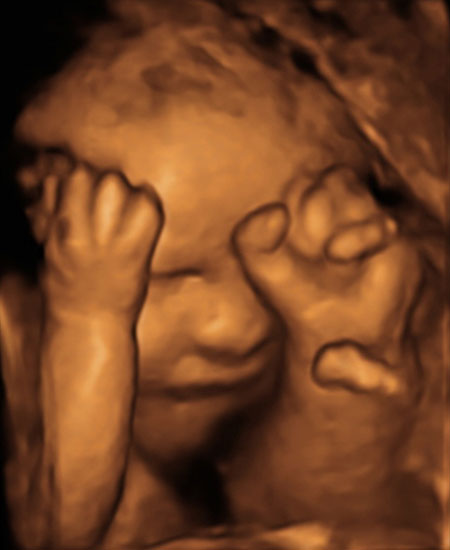
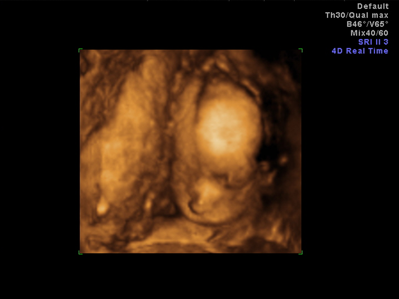
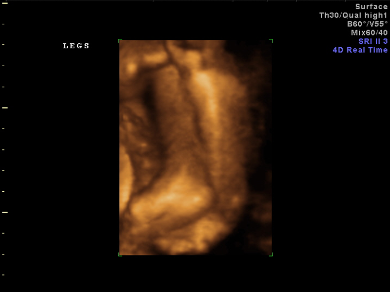
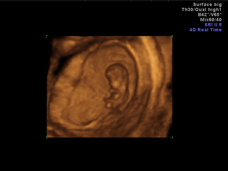
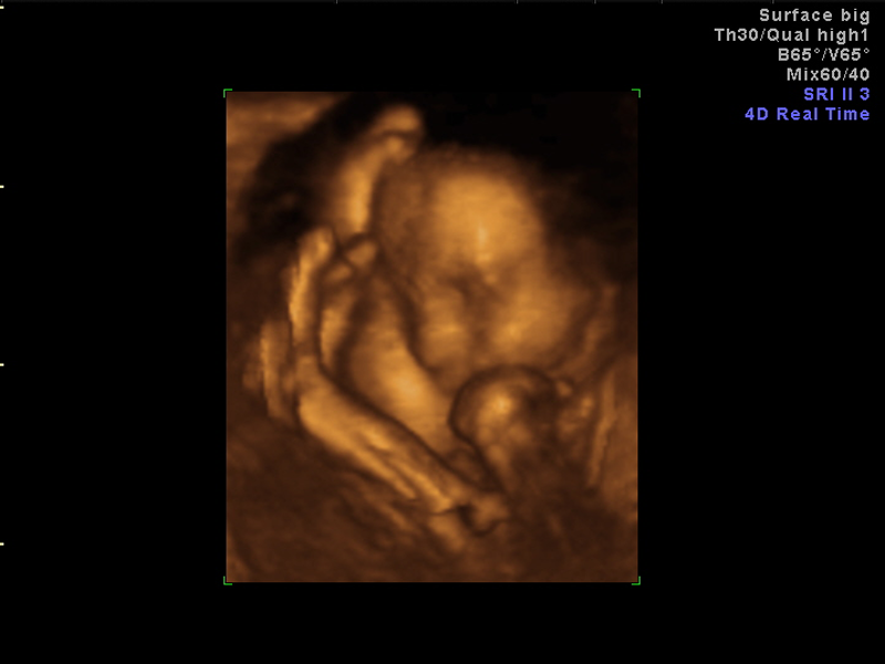
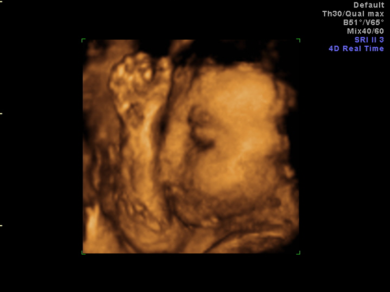
This technique can provide excellent life-like images of a baby in utero. It can be used to further assess the baby for fetal abnormalities but mostly is used to reassure parents that their baby looks normal.
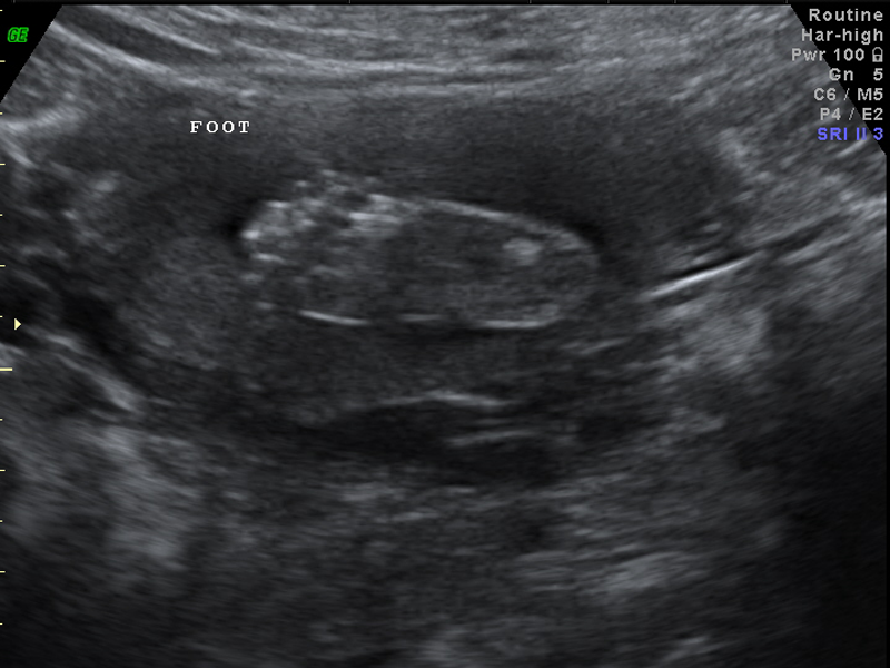
At this stage, however, the most important part of the examination to assess the fetus still depends on a thorough 2 dimensional grey-scale examination which allows a more careful examination of the internal structures of the baby.
4D ultrasound is used at all the obstetric ultrasound examinations. Excellent and life like images can be obtained at all stages of pregnancy. However, the images obtained are not always as cute as one would wish.
All pictures obtained depend very much on the position of the baby and of the amount of amniotic fluid.
At the 12 week NT examination the baby can be seen completely but will have a big head and small body. Sometimes the arms and legs will not look complete. The young baby before 20 weeks will have a very thin face and at times will look very much like ET.
From 26-28 weeks onward the baby will have more tissue between the skin and bones of the face and will look much more babyish.
Share precious moments with your baby
Our state-of-the-art digital equipment produces top-quality 3D and 4D ultrasound images. So not only do you get an on-the-spot diagnosis and assessment, but also, you get a high-quality image of your baby to treasure and share with those closest to you.
To give you further reassurance, we provide copies of reports, printed images, CDs of images and DVDs of your scan; whatever you need for your comfort and peace of mind.
Click here to learn more about getting a 3D/4D precious moment of your own.
